

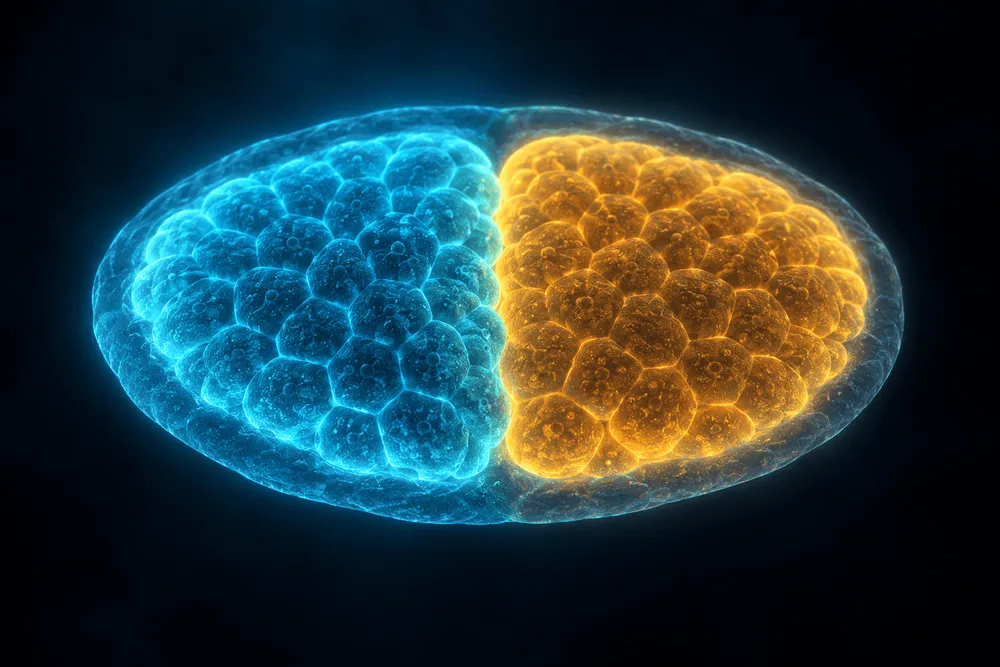
A paper published in Nature Neuroscience on September 18, 2026 showed that the front and back of the brain originate from distinct progenitor cells from a very early stage of embryonic development, and begin forming in parallel.
That same day, Stanford Medicine's press release headline stated that "the human brain is two separate organs," and corresponding author Kyle M. Loh told an interviewer, "At the very least, there are two separate organs."
However, the paper's abstract used the verb "postulate"—a hedged, tentative phrasing—and referred to the brain in the singular as a "composite organ."
Over the following five days, coverage of the same study ranged from flat assertion to careful verification, cautious hedging, and outright denial. Depending on which article a reader encountered first, they could walk away with opposite conclusions.
So what exactly did this paper show?
Not a Branching Path, But Two Parallel Origins From the Start
The paper's title is "Two parallel neural ectoderm progenitors contribute to the developing brain" (DOI 10.1038/s41593-026-02433-7). The corresponding author is Kyle M. Loh, associate professor of developmental biology at Stanford University; co-first authors are graduate students Rayyan Jokhai and Carolyn Dundes.
The research centers on gastrulation—the period when the embryo's basic body plan, including the front-to-back axis, begins to take shape.
At this stage, anterior neural ectoderm expressing the gene Otx2 and posterior neural ectoderm expressing Gbx2 emerge almost simultaneously. The former develops into the forebrain and midbrain; the latter into the hindbrain, which forms the bulk of the brainstem.
The traditional textbook model held that the neural ectoderm—the tissue that gives rise to the brain—first forms as a single continuous sheet, which is only later subdivided into anterior and posterior regions.
This paper challenges that sequence. Rather than posterior progenitors splitting off later from anterior ones, the evidence presented shows that the two arise separately from an early stage of development.
That said, the authors themselves do not rule out the possibility that, for a very brief window even earlier, a common progenitor capable of generating both anterior and posterior tissue may have existed.
Verification in mouse embryos used a technique that labels only cells present at a specific time point and tracks their descendants.
Mice carrying a CreER enzyme active in Gbx2-expressing cells were crossed with reporter mice that begin producing the red fluorescent protein tdTomato once Cre is activated.
Because CreER only translocates into the nucleus and becomes active after drug administration, a single dose of 4-hydroxytamoxifen at embryonic day E7.5 labels cells that were posterior neural ectoderm at that moment, within roughly a 12-hour labeling window.
The researchers then tracked where the descendants of these labeled cells migrated.
The result: descendants of the labeled cells remained almost entirely confined to the hindbrain from E8.5 through E18.5, with virtually no appearance in the forebrain or midbrain.
Tracking at single-cell resolution provided a more detailed breakdown.
Of 494 neural ectoderm clusters derived from 16 independent embryos, 62.96% were found exclusively in the forebrain/midbrain, and 32.59% exclusively in the hindbrain. Combined, 95.55% fell cleanly into one region or the other.
The remaining roughly 4% could not be classified as belonging exclusively to either region. Whether this reflects a limitation of the experimental system or a genuine biological phenomenon—such as cell migration near the boundary—remains unknown.
Ars Technica rounded this result to "96% of cases" and noted that the reason it isn't 100% remains unresolved.
For humans, the experiments were conducted in culture dishes rather than living embryos.
Anterior and posterior neural ectoderm were each generated from human pluripotent stem cells, then exposed to signals that would normally promote the opposite fate.
If cell fate were determined solely by the surrounding environment at that moment, anterior cells exposed to posteriorizing signals should have been able to convert into hindbrain-type cells.
But in practice, no such conversion occurred.
The authors interpret this as "lineage commitment"—a state in which the capacity to switch to a different cell lineage has been substantially restricted.
One piece of supporting evidence involves chromatin: how DNA is packaged, and which genes are rendered accessible as a result.
In anterior and posterior progenitors, the regions surrounding genes each lineage would later use were already open, while regions needed for the opposite fate were closed.
In Stanford Medicine's press release, Loh stated: "We show for the first time that the front of the brain arises from a completely different progenitor than the back."
Where Did the "Two Organs" Framing Come From?
While the paper's abstract used the cautious verb "postulate" and the singular "composite organ," the university's press release used "is" and the plural "organs."
The paper's abstract concludes: "We postulate the brain is a composite organ emanating from two lineage-restricted progenitors."
By contrast, the headline of Stanford Medicine's press release, published September 18, 2026, read: "Human brain is two separate organs, Stanford Medicine-led research finds."
The assertive framing wasn't limited to the headline.
The body of the press release also used strong language, stating that the research "reveals the brain is actually two separate organs" and "overturns conventional thinking."
The more hedged phrase "may actually be" didn't appear in a headline until three days later, on September 21, when ScienceDaily covered the same press release.
Looking at the precedents cited in the paper's discussion section helps clarify what "composite organ" actually means.
The heart, for example, is built from distinct progenitor populations—the first and second heart fields—yet it is not typically called "two organs" on that basis.
What the paper proposes is a framework in which a single organ forms from two distinct starting points—not a claim that the fully formed brain is anatomically or functionally split into two independent organs.
In Stanford Medicine's press release, Jokhai touched on the possibility that roughly 500 million years ago the two systems were independent nervous systems, but added that "now they function largely as one."
That said, it would also be inaccurate to say the "two organs" phrasing was invented solely by university communications staff.
In an interview published the same day, September 18, on Nautilus, Loh was asked about the unresolved origin of the cerebellum and the possibility of a third developmental pathway. He responded: "We don't have the data to support or refute that. But at the very least, there are two separate organs."
In other words, a gap between the cautious language of the paper itself and the language the author used in a public-facing setting had already emerged on the very same day.
That said, an interview involves spoken explanation. Using strong language there does not necessarily mean the author was retracting the qualifications stated in the peer-reviewed paper.
Coverage in Japanese was likewise inconsistent.
Nazology, on September 21, framed it as "what we call the 'brain' was actually a combination of two different organs," and Tech-i Life Science, on September 22, adopted similarly assertive language: "discovered that the human brain is two separate organs."
By contrast, CodeBeat News—the earliest Japanese-language article confirmed—published on September 20 that "the 'two organs' framing is the researchers' interpretation used to explain their results," and explained that the paper itself uses the term "composite organ."
Four Claims, Not Equally Well Supported
Some of the figures below are also drawn from the peer-reviewed preprint of the same study (bioRxiv, July 5, 2025).
The published version in Nature Neuroscience is subscription-only, and details may have changed through peer review. For that reason, confidence in the claims shouldn't hinge on decimal-point figures alone—the overall structure of the study matters more.
Laying out the four major claims alongside their type of evidence, experimental conditions, and how the authors themselves framed them reveals that their levels of confidence differ.
| Claim | Type of evidence | Sample and conditions | Authors' framing |
|---|---|---|---|
| Anterior and posterior neural ectoderm arise in parallel, with descendants remaining separate | In vivo lineage tracing in mouse embryos | Gbx2-CreER × tdTomato reporter. Administered at E7.5 with roughly a 12-hour labeling window; tracked from E8.5 through E18.5. Single-cell labeling covered 494 clusters from 16 embryos | Reported that descendants remained largely confined to the hindbrain |
| "Lineage commitment" prevents conversion to the opposite fate | Culture experiments using human pluripotent stem cells | Anterior and posterior cells each exposed to signals promoting the opposite fate | Explicitly stated it remains undetermined whether the same lineage commitment holds true in vivo |
| Chromatin state reflects future fate | OmniATAC-seq and RNA-seq on human pluripotent stem cells | Sequencing data deposited in GEO as GSE286214 | Reported differing chromatin accessibility between anterior and posterior cells |
| The two lineages have been conserved for over 500 million years | In situ staining in chicken, zebrafish, acorn worms, and others | According to the figure legend, a single embryo was analyzed per species | Suggests the possibility of evolutionary conservation |
What was directly demonstrated in mouse embryos is fate tracking—that is, which region a cell's descendants end up in.
By contrast, the evidence for lineage commitment—the idea that once anterior or posterior fate is set, it cannot be switched—comes from culture experiments using human pluripotent stem cells.
The authors themselves state explicitly in their discussion that whether anterior and posterior neural ectoderm are similarly lineage-committed in vivo remains unresolved.
Directly testing this would require transplanting labeled cells to a different location in the gastrulating embryo, which they note is technically difficult.
Generalizing "once a cell's fate is set, it cannot switch to another fate" to the whole organism therefore means extending a result obtained in a culture dish to actual embryonic development.
The authors also raise an important possibility that current experiments cannot rule out.
Between the time the ectoderm forms (roughly E6.75–E7.0) and the time anterior and posterior neural ectoderm become distinguishable (roughly E7.5), a "pan-brain progenitor" capable of generating the entire brain may have existed for a very brief window.
Whether a common ancestral cell for both anterior and posterior lineages existed before the point at which this study's tracking begins remains unknown.
The evolutionary claims also vary in specificity.
The paper states the common ancestor existed roughly 550–600 million years ago; Stanford Medicine's press release says "more than 550 million years ago"; and Jokhai's comment cited roughly 500 million years ago.
Regarding the multi-species in situ staining used as evidence for evolutionary conservation, the paper's figure legend indicates that a single embryo was used per species.
While the evidence points to the possibility that the divergence between anterior and posterior developmental pathways has very ancient origins, it isn't strong enough to pin down a precise date.
What This Study Adds to a 1952 Model
The question of when different brain regions diverge has been debated for more than 70 years.
In 1952, Nieuwkoop proposed a model in which the neural ectoderm possesses broad developmental potential, capable of giving rise to the forebrain, midbrain, and hindbrain alike.
Some current methods for generating brain cells from human stem cells implicitly rely on this idea.
The approach is to first generate neural progenitor cells, then apply various signals afterward to specify anterior or posterior identity.
Meanwhile, fate maps constructed since the latter half of the 20th century in zebrafish, frogs, chickens, and mice suggested that, even at the gastrula stage, neural ectoderm cells already tend toward particular brain regions.
However, fate maps have a limitation.
Simply tracking cells left in their original location cannot distinguish whether a cell "acquired that fate because of its local environment" or whether it "had already lost the ability to choose a different fate."
As a result, a model in which cells start from a common progenitor and only later diverge in response to surrounding signals could not be ruled out by fate maps alone.
One key contribution of this study is an experimental distinction between being "fated" (destined to become something) and "committed" (unable to switch to a different fate).
By generating anterior and posterior progenitors from human pluripotent stem cells and exposing each to signals promoting the opposite fate, the researchers tested this distinction directly.
That said, the idea that anterior and posterior regions diverge early is not itself new.
CodeBeat News points out that the boundary marked by Otx2 and Gbx2 was already known from earlier studies published in 2001 and 2011.
The author himself has spoken about the debate the field went through before accepting this study's findings.
In an interview, Loh said, "It was a real struggle to get this published, because it was widely believed that what we found wasn't correct," and recalled that at a conference several years ago, the head of a developmental biology department stood up and declared, "This is completely wrong."
Notably, the preprint's title was "Two parallel lineage-committed progenitors contribute to the developing brain," but the word "lineage-committed" was dropped from the title in the published version.
There isn't enough evidence to conclude this reflects a reduction in confidence through peer review—but the title change itself is confirmed.
The Same Study, Told in Opposite Ways Within Five Days
Coverage of this paper didn't simply grow more cautious over time, nor did it simply grow more assertive.
From the very first day, strong assertions and cautious hedging existed side by side, and differing interpretations continued to spread afterward.
Here are eight confirmed articles, listed chronologically:
| Date | Outlet | Headline/claim | Strength of framing |
|---|---|---|---|
| 2026-09-18 | Stanford Medicine | Human brain is two separate organs | Assertive, though with some hedging in the body |
| 2026-09-18 | Neuroscience News | The brain is two separate organs joined by evolution | Assertive |
| 2026-09-18 | Nautilus | You have two brains, not one | Assertive; author interview |
| 2026-09-20 | CodeBeat News | What's behind the "two brains" theory | Verification |
| 2026-09-21 | ScienceDaily | Human brain may actually be two separate organs | Hedged |
| 2026-09-21 | Nazology | A combination of two different organs | Leaning assertive |
| 2026-09-22 | Tech-i Life Science | Discovered to be two separate organs | Assertive |
| 2026-09-23 | Ars Technica | Contrary to some reports, you don't have two brains | Denial |
This wasn't a simple case of the university's press release using strong language that spread through outlets over time before a specialist publication finally corrected it.
Three assertive articles appeared on day one alone, one of which was an interview with the author himself.
Meanwhile, articles that carefully scrutinized the content also appeared early. As far as could be confirmed, the Japanese outlet CodeBeat News published a verification article on September 20—three days before Ars Technica published its skeptical rebuttal in the English-language press.
In other words, which conclusion a reader walked away with depended less on publication timing overall and more on which specific article they happened to read first.
The pushback deserves closer examination too.
On September 23, Ars Technica published "Contrary to some reports, you don't have two brains," in which John Timmer criticized the framing: "You don't have two brains. That's an extreme reading of what this paper actually found."
Timmer is a senior science editor at the outlet; according to his Muck Rack profile, he holds a Ph.D. in molecular and cell biology and spent over a decade doing research in genetics and developmental biology.
The article also traces the origin of the "two brains" framing to Stanford Medicine's press release.
However, on this point, corresponding author Loh himself used the phrase "two separate organs" in an interview published the same day, September 18.
That makes it difficult to attribute the framing solely to university communications.
That said, this represents the assessment of a single Ars Technica reporter; as far as could be confirmed, multiple independent developmental biologists have not published similar critiques of this study.
The eight articles listed here also do not represent an exhaustive survey of everything published during this period.
A Potential Path to Growing Neurons Involved in Swallowing
Setting aside the question of whether the brain is "one organ or two," the practical significance of this study becomes easier to see.
The authors developed a culture protocol starting from posterior neural ectoderm, and used it to generate motor neurons from human pluripotent stem cells with characteristics specific to hindbrain rhombomeres 5/6.
These cells express genes including HOXA1, HOXA3, PHOX2A, and PHOX2B, and in the body are involved in functions such as swallowing.
Hindbrain-derived motor neurons have historically been among the more difficult cell types to generate in culture dishes.
This result suggests part of the reason may lie in the starting point of the culture protocol itself.
Jokhai stated: "The field has been trying to coax forebrain and midbrain progenitors into becoming hindbrain cells, but our research shows that this doesn't work."
In other words, if the wrong progenitor cell is chosen as a starting point, subsequent signaling is unlikely to reach the target cell type.
Such cells are also relevant to research on spinal muscular atrophy (SMA) and ALS (amyotrophic lateral sclerosis).
Both are designated intractable diseases in Japan, and as they progress, swallowing function can decline, sometimes leading to complications such as aspiration pneumonia.
If motor neurons involved in swallowing can be recreated in a culture dish, it may become possible to study, in detail and outside the patient's body, the process by which specific cell types are selectively lost.
The fact that the Spinal Muscular Atrophy Foundation is among the study's funding sources also points to this intended application.
Meanwhile, unresolved questions remain, and are explicitly acknowledged both in the paper and by the authors themselves.
Demonstrating that anterior and posterior neural ectoderm are truly lineage-committed in vivo would require experiments such as transplanting labeled cells to different locations—something the authors describe as technically difficult.
As for which lineage the cerebellum originates from, Loh answered that "we don't have the data to support or refute" any particular answer, leaving open the possibility of a third developmental pathway.
And the roughly 4% of cells that could not be classified as belonging exclusively to either the anterior or posterior lineage remains unexplained.
At this stage, what the study demonstrated relatively clearly can be narrowed down.
Under these culture conditions, once cells diverged early in development into anterior and posterior neural ectoderm, switching them to the opposite fate afterward proved difficult.
This research suggests that anyone trying to generate hindbrain-derived cells from human pluripotent stem cells may need to reconsider the conventional approach of starting from forebrain-type progenitors altogether.