

When neuroscientists study human brain development or disease mechanisms, the thickest wall they face is the simple fact that their subject is a living human being. Opening a skull to directly manipulate the living neural circuits of a healthy or diseased human brain, tracking molecular and cellular interactions step by step, is neither ethically nor physically permissible. Researchers have long relied on three-dimensional "cerebral organoids" grown in petri dishes from pluripotent stem cells, but tissue in a test tube has no blood vessels to carry blood, no sensory input, and no muscles to drive as output.
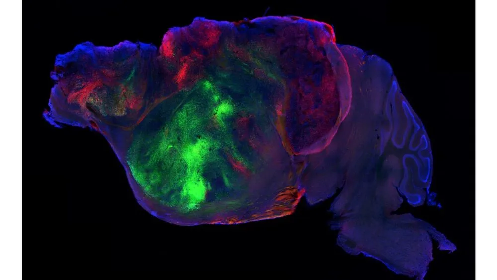
A research team led by Professor Sergiu Pașca at Stanford University School of Medicine has published results tackling this longstanding constraint in an open-access paper in the British scientific journal Nature. The team engineered a mouse strain in which most of the cerebral cortex and hippocampus are eliminated during early development through genetic modification, then transplanted cortical organoids derived from human stem cells into the resulting void. The transplanted human tissue expanded rapidly within the skull, growing to occupy over 90% of the cortical space within a few months after birth. An accompanying commentary in Nature (Nature News d41586-026-02912-8) described these mice as achieving "the most extensive functional integration of human brain cells into an animal reported to date," and the researchers have named them "xenocortical mice."
However, it would be scientifically incorrect to interpret this finding as "the creation of mice with human intelligence or consciousness." The human tissue filling the skull remains in an extremely immature state, closer to a mid-gestation fetal brain, and lacks the mature six-layer structure of the fully developed cerebral cortex. Furthermore, whether the partial behavioral recovery observed in maze tests stems from computations performed by the human cells themselves, or is merely a secondary phenomenon in which the mouse's existing circuits respond to physical stimulation, has not yet been proven.
Genetic Modification to Eliminate Competition, and Human Organoid Transplantation Into the "Void"
This is not the first attempt to transplant cerebral organoids into an animal brain. In 2022, Professor Pașca's laboratory transplanted human cerebral cortical organoids into the somatosensory cortex of newborn rats and demonstrated that human neurons responded electrically to stimulation of the rat's whiskers. However, that earlier study faced an insurmountable barrier.
Rodents and humans differ fundamentally in the speed of nervous system development. While rat brain cells proliferate rapidly and weave together robust neural circuits within weeks, human neurons require months to years to mature. As a result, the host rat's own neural circuits quickly occupied the physical space and synaptic connection targets within the brain, forcing the human organoid to stop growing once it was confined to roughly one-third of the cortical hemisphere. This asymmetry in developmental speed created fierce interspecies competition for both space and connection targets.
データを表で見る
| Maximum Occupancy Rate (%) | |
|---|---|
| 2022 Prior Study (Normal Rat) | 33 |
| 2026 Current Study (Apallial Mouse) | 90 |
As this chart shows, the presence or absence of competing host cortical cells causes nearly a threefold difference in the volume that human neural tissue can occupy within the brain. The team arrived at the idea of eliminating this competition altogether.
The research team developed an immunodeficient mouse strain in which tissue differentiating from the dorsal and medial cerebral pallium is specifically eliminated early in development through genetic engineering. This mouse carries the genotype Emx1-cre; Esco2 fl/fl; Prkdc scid/scid, with genes involved in maintaining division of cerebral cortical cells selectively disrupted. The team named this condition the "apallial mouse."
In the apallial mouse's brain, approximately 98% of the neocortex and hippocampus is lost during development. Whole-brain MRI imaging revealed roughly a 50% reduction in total brain tissue volume compared to untreated wild-type controls. However, subcortical structures such as the basal ganglia, thalamus, cerebellum, and brainstem remain almost entirely intact. This vast void is prepared within the skull shortly after birth, and cortical organoids (hCOs) derived from human iPS cells are then injected into it.
The transplanted human organoid began expanding in the unobstructed space, drawing nutrients from the mouse's bloodstream. Although the paper and available published materials do not provide absolute volume figures for before-and-after comparison, by roughly two to three months after injection the human tissue had expanded approximately fivefold in volume, filling over 90% of the cranial space that would normally be occupied by the mouse's own cerebral cortex.
Axons Reaching the Spinal Cord and the Emergence of Von Economo-Like Neurons
The human cells filling the skull did not simply proliferate as an inert mass. Blood vessels from the host mouse's circulatory system rapidly invaded the tissue, and a microvascular network autonomously formed, supplying oxygen and glucose. Whereas conventional static petri-dish cultures were limited to a few millimeters in diameter due to necrosis at the tissue core, the living blood supply allowed the tissue to survive deep into its interior.
Measurements of neural activity within the tissue using two-photon calcium imaging and electrophysiological recording revealed that the human neuron population exhibited spontaneous electrical activity. Moreover, the firing patterns were not disordered noise but displayed coordinated synchronization patterns characteristic of developing neural circuits.
The most striking anatomical feature was the path taken by axons extended by human neurons. Nerve fibers traced with fluorescent labeling crossed the boundary of the transplant region and penetrated deep into the mouse's own brain tissue. The fibers passed through the mouse's thalamus and basal ganglia, and even through the brainstem, with some crossing past the neck's nervous system to reach deep into the spinal cord. Layer 5 extratelencephalic-projecting neurons (L5-ET neurons), which are responsible for long-distance projections, differentiated within the tissue, demonstrating that the wiring program native to the human brain was activated even in this ex vivo environment.
What surprised the research team even further was the discovery, within collected tissue sections, of unusual elongated spindle-shaped cells known as "von Economo-like neurons." Unlike typical cortical pyramidal cells, which have dendrites spreading in a fan shape, this cell population has a distinctive morphology with a single thick polar process extending both upward and downward.
Von Economo neurons are specialized large cells found almost exclusively in the anterior cingulate cortex and anterior insular cortex of large mammals with highly advanced social behavior, such as humans, great apes, cetaceans, and elephants. They are known to be selectively lost in the early stages of frontotemporal dementia (FTD), and have drawn attention as a cell type that may hold the key to psychiatric disorders and cognitive changes. However, reliably producing these cells in flat culture dishes or standard organoid cultures in vitro has proven extremely difficult. This cell population, which had not been observed even in the 2022 rat transplantation study, spontaneously emerged within the skull of the apallial mice in this new study.
Why did this rare cell type fail to develop in culture dishes but appear only inside the mouse skull? In an interview with ScienceAlert, Professor Pașca noted that the cause remains unknown, but offered two hypotheses.
First, the physical connection of human neuron axons to distant targets such as the mouse spinal cord may have allowed the cells to receive retrograde signals released from the target tissue. Second, growth factors present in the living host's bloodstream and surrounding interstitial fluid, direct interaction with vascular endothelial cells, or spontaneous electrical activity itself may have functioned as microenvironmental signals that cannot be replicated in vitro. However, these remain working hypotheses derived from observed phenomena, and which factor drove the cell fate decision has not been experimentally proven.
Above all, this human tissue remains structurally far from a complete cerebral cortex. Even approximately six months after transplantation, the tissue's stage of differentiation remained at the immature level of the human mid-gestation period (roughly 18 to 24 weeks). The orderly six-layer structure (lamination) seen in the adult human brain had not formed, and cells remained mixed together in a coarse mass.
The Gap Behavior Experiments Exposed Between "Functional Recovery" and "Causality"
What changes appear in an animal's behavior when human neural tissue is implanted into the skull of a mouse that has lost its cerebral cortex and hippocampus? The research team conducted multiple behavioral tests across three groups: normal control mice, apallial mice without human cell transplants, and xenocortical mice with transplanted human tissue.
The primary evaluation metric was the standard "Y-maze test," which measures working memory (short-term memory) in mice. A mouse placed in a Y-shaped track has an innate tendency to preferentially explore an arm it has not just explored (spontaneous alternation behavior). This alternation rate will not increase unless the mouse remembers which passage it entered most recently.
| Group Classification | Cerebral Cortex and Hippocampus Status | Presence of Human Tissue | Y-Maze Test (Working Memory) | CatWalk Gait Analysis (Fine Motor Coordination) | Free Behavior Analysis (MoSeq) | Key Caveat for Interpretation |
|---|---|---|---|---|---|---|
| Control | Normal | None | Success rate significantly above chance | Normal limb placement and smooth gait | Forms normal spontaneous behavior pattern group | Baseline data from wild-type mice |
| Apallial | ~98% deficit from early development | None | Equivalent to chance level (50%), memory impairment | Severe motor coordination impairment, abnormal foot placement | Behavior significantly deviates from normal pattern | Direct impairment due to absence of cortex and hippocampus |
| Xenocortical | Human tissue reconstituted in mouse's deficient region | Yes (over 90% of volume) | Recovered to level significantly above chance | Only partial coordination improvement, deficits remain | Distinct pattern positioned between control and apallial groups | Whether human neurons are directly involved or produce an indirect effect remains unidentified |
Apallial mice lacking cortex and hippocampus could not surpass chance level (the probability of a random guess) in the Y-maze, showing clear short-term memory impairment. Gait analysis using a CatWalk apparatus also recorded severe motor impairment, with disrupted coordination among the limbs.
In contrast, xenocortical mice with established human tissue showed alternation behavior significantly exceeding chance level in the Y-maze test. The lost exploratory behavior associated with short-term memory had recovered with statistical significance.
Three-dimensional behavioral analysis using machine learning (MoSeq), which classified hundreds of thousands of frames of free behavior, also showed that the behavioral sequences of xenocortical mice diverged from the pathological pattern of apallial mice, tracing a distinctive trajectory positioned right between the control and apallial groups.
データを表で見る
| Alternation Rate Level (%) | |
|---|---|
| Chance Level (Theoretical) | 50 |
| Apallial Mouse (Deficit) | 50 |
| Xenocortical Mouse (Post-Transplant) | 63 |
| Normal Control Mouse | 68 |
Taken in isolation, this result invites an appealing narrative: "human brain cells revived a mouse's memory." However, Professor Pașca's own comments to ScienceAlert were notably restrained.
"This is an interesting result, particularly given that apallial mice did not show the same capability. But we should be cautious about concluding from this fact alone that human neurons are directly responsible for that behavior."
Professor Adeel Razi, who studies neuroimaging and brain networks at Monash University in Australia, issued a similar warning through the Australian Science Media Centre. Professor Razi points out that "functional integration being confirmed does not mean functional equivalence." It remains extremely difficult, within the current experimental design, to completely rule out the possibility that electrical activity generated within the skull provided nonspecific arousal stimulation or nutritional support to existing circuits in the mouse's hypothalamus or brainstem, with the mouse's own remaining tissue driving the behavioral improvement. Whether human cells processed specific information and sent deliberate command signals to the mouse's muscles must await future causal verification.
The Scope and Structural Gaps of a Disease-Research Platform
This xenocortical mouse model was developed not as bioengineering to enhance cognition, but as an "experimental platform" for reproducing, at the level of the whole organism, how human brain diseases develop.
In the paper, the research team presents one proof of concept as a disease model: modeling hypoxic encephalopathy resulting from conditions such as perinatal asphyxia. When xenocortical mice were exposed to a hypoxic environment for a set period, host-derived microglia and astrocytes infiltrated the transplanted human tissue and expressed specific hypoxic stress markers. Furthermore, clear impairment in gait coordination appeared in CatWalk testing.
With conventional petri-dish culture, it was impossible to measure how immune cell infiltration via blood vessels occurs, or how hypoxic stress propagates to affect whole-body motor function. Using xenocortical mice demonstrated that it is possible to measure "the pathological response of human neural tissue coupled with a living circulatory system."
Professor Pașca cites the following three areas as the primary future applications.
The first is the aforementioned frontotemporal dementia (FTD). By generating cortical tissue from patient-derived iPS cells and growing it inside the mouse skull, researchers can track within the living organism why the von Economo-like neurons that emerge die off before other cell types—the mechanism behind their selective vulnerability.
The second is research into treatment-resistant epilepsy. This involves elucidating what kind of abnormal synchronous firing human neural circuits carrying specific genetic mutations generate, and how that activity spreads through the brainstem to cause whole-body seizures, as well as screening candidate anti-epileptic compounds in vivo.
The third is neurodevelopmental disorders, including autism spectrum disorder (ASD) and schizophrenia. There is potential to quantitatively and longitudinally observe how abnormalities in synaptic pruning and circuit connectivity specific to the human cerebral cortex are reflected in an animal's behavioral sequences.
However, this platform has a decisive biological shortcoming. As Professor Pașca himself acknowledges, GABAergic neurons responsible for inhibitory neurotransmission do not autonomously differentiate within the transplanted tissue. Because cerebral cortical organoids primarily generate excitatory glutamatergic neurons of the dorsal cortex, they lack the inhibitory interneurons that would normally migrate in from the ventral ganglionic eminence.
Neural tissue lacking inhibitory circuits exists in a significantly imbalanced state between excitation and inhibition. Combined with the absence of the six-layer laminar structure, Professor Pașca emphasizes: "This is not an all-purpose universal system. It is a specialized tool designed to answer extremely limited, specific disease questions."
The Boundaries of Animal Identity and Laboratory Ethical Standards
Attempting to grow human brain cells at scale within an animal's skull and connect them to the central nervous system inevitably raises profound bioethical questions. When over 90% of a mouse's brain has been replaced with human tissue, is that organism still simply a "laboratory mouse," or has it transformed into something requiring different ethical consideration?
The research team incorporated this ethical challenge not as an afterthought following the study's completion, but from the very planning stages of the experiment. In addition to review by the standard animal experimentation committee, an independent ethics review board was formed, composed of Stanford bioethicists, external neuroscientists, legal scholars, and patient advocacy representatives, and this board conducted ongoing evaluation in parallel with the progress of the experiments.
Two central concerns were examined. One is the basic principle of animal welfare: whether the suffering experienced by the experimental animals is proportionate to the medical knowledge gained from the research, and whether alternative methods exist. The other is the concern that, as more complex human neural tissue becomes connected to an animal's nervous system, unforeseen "emergent properties" (emergent cognition or emotion) might arise.
Professor Pașca states that he took the latter possibility seriously and closely monitored the mice's behavior and physiological responses throughout the entire experimental period. He further stated:
"The most important point is that these remain mice. They have a mouse nervous system, mouse sensory organs, and mouse subcortical structures. What is unusual is that the majority of the cortical tissue is human-derived, and that these cells are functionally connected to the mouse's nervous system."
At the same time, the research team and bioethicists point to "the ethical cost of failing to develop better disease models." Neurological and psychiatric disorders affect nearly one in five people worldwide, and for many of these conditions, both the fundamental molecular mechanisms and effective drug treatments remain elusive. Conventional animal models cannot reproduce pathology specific to the human cerebral cortex, and isolated cultured cells cannot be used to verify circuit-level disruption at the organism level. It was to break through this impasse that the xenocortical mouse was designed as an experimental testbed.
This achievement represents a technical milestone in successfully engrafting human tissue at scale within a living animal's brain—it does not represent the completion of a clinical treatment or drug for human use. The human cells within the skull have not shed their fetal-stage immaturity, and the fundamental question of whether the memory recovery observed in the maze stems from logical computation by human neurons remains unanswered by anyone. What does a human neuron learn, and what does it drive, within the complex environment of a living body? What science has obtained is a fragile, limited new observation window into that vast mystery.