

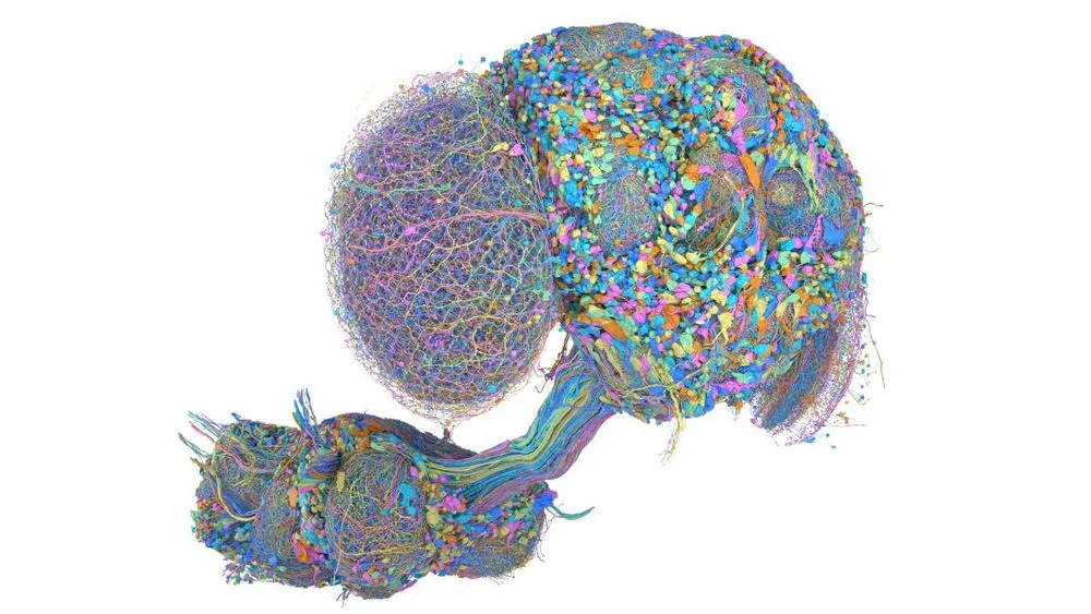
When an animal perceives the world and acts upon it, countless neurons inside its brain exchange signals through synapses. The map that underlies our understanding of this information flow is the connectome—a wiring diagram of neural circuits. Just as you cannot fundamentally repair an electronic device without its circuit diagram, you cannot truly understand memory, learning, or the mechanisms of neurological disease without knowing the brain's structural foundation.
However, mapping the human brain—with its roughly 86 billion neurons—at synaptic resolution remains beyond the reach of current technology. Cutting out just one cubic millimeter of human cerebral cortex yields tens of thousands of neurons and hundreds of millions of synapses, generating petabytes of data. For this reason, neuroscientists have turned to far smaller model organisms, mapping their nervous systems from end to end in search of universal operating principles governing brains as systems.
The fruit fly (Drosophila melanogaster) is the prime example. Ever since Seymour Benzer founded behavioral genetics in the 1970s, this insect—with its short life cycle and stereotyped behaviors—has driven discoveries that led to multiple Nobel Prizes. Yet even its tiny brain is no simple matter to wire-map. Brain tissue must be sliced into millions of ultra-thin sections, photographed under an electron microscope, and reconstructed three-dimensionally. For years, researchers were burdened by the tedious, seemingly endless manual work of tracing cell boundaries. Today, machine learning algorithms are fundamentally transforming this process.
AI Fills In Electron Microscope Images, Surpassing the Limits of Manual Tracing
To break through the limitations of manual labor, a team at Google Research turned to artificial intelligence. They developed a method using a convolutional neural network called "Flood-filling networks (FFN)," which identifies pixels belonging to the same object in two-dimensional electron microscope images and automatically traces and reconstructs the three-dimensional structure of neurons.
The first step in building a connectome is slicing brain tissue into ultra-thin sections. The fly's brain is embedded in resin and cut into slices tens of nanometers thick using a diamond knife. These slices are then photographed under an electron microscope and digitally reassembled into a three-dimensional block on a computer. The biggest challenge here is separating the countless intertwined neuronal branches from one another.
Conventional image segmentation algorithms separated neurons by predicting the boundaries of cell membranes. But in noisy electron microscope images, tiny errors tend to accumulate. Cell membrane boundaries are often blurred by uneven staining, and it's not uncommon to mistake the outlines of mitochondria or synaptic vesicles within the image for cell boundaries. A single small gap can lead to a false connection with a different neuron, and if an axon—which branches thousands of times—is mistraced at even one point, the entire circuit can collapse.
FFN takes a different approach: rather than segmenting the entire image at once, it starts from a single point (a seed) and expands the network, filling in regions belonging to the same cell. This method allows dendrites with complex shapes to be traced with high accuracy. The process resembles how human vision follows the lines of a maze, preserving local shape context throughout the reconstruction.
Back in 2020, the team published a hemibrain map of a female fruit fly. That effort recorded 25,000 neurons and 21 million synaptic connections. Years later, the latest reconstruction system, called "PATHFINDER," improved both the speed and accuracy of reconstruction by incorporating synthetic neurons into its training data. AI produces a draft, and a team of experts at the HHMI Janelia Research Campus performs final verification and error correction. This collaborative process between humans and machines has made it possible to produce brain maps of a scale once estimated to take decades, within a realistic budget and timeframe.
An Uninterrupted Map of 125 Million Synapses, From Brain to Ventral Nerve Cord
In a study published in the journal Cell in September 2026, an international team including HHMI Janelia and the University of Cambridge released a connectome covering the entire central nervous system of a male fruit fly (DOI: 10.1016/j.cell.2026.08.015). This map includes not only the brain but also the ventral nerve cord, the equivalent of a spinal cord, encompassing approximately 166,000 neurons and 125 million synaptic connections. In terms of neuron count within a single individual, this is the largest completed brain map to date.
The key advance of this research lies in the fact that the brain and ventral nerve cord were mapped while remaining connected. Previous connectome studies had been limited to circuit diagrams isolated to specific regions. But this new map allows researchers to trace, without interruption, the entire pathway—from the brain capturing external visual and olfactory stimuli, through signals descending via the neck to the ventral nerve cord, to their conversion into motor output that moves the legs and wings.
Inside the fruit fly's brain lie the mushroom body, a center for memory and learning, and the central complex, a hub for spatial navigation. Previous research had only fragmentarily revealed the internal wiring of each functional region. But this complete central nervous system map now reveals the physical pathway by which an odor memory formed in the mushroom body travels to the central complex, integrates with directional sense, and transforms into a concrete motor command to move toward the source of the smell.
The insect's ventral nerve cord is not merely a conduit for commands from the brain. It is a center that autonomously governs the generation of complex motor patterns, such as walking reflexes and the timing of wingbeats. The significance of being able to analyze the entire flow—from sensory input to behavioral output—as a single continuous circuit is substantial. When a particular neuron fires, researchers can now confirm, as fact rather than inference, exactly which muscle movement it ultimately produces. This can be traced all the way through on a single wiring diagram, without having to splice in data from a different individual midway.
データを表で見る
| Number of synapses (millions) | |
|---|---|
| Female hemibrain (2020) | 21 |
| Male full CNS (current) | 125 |
As the chart above shows, expanding the scope from the hemibrain to the entire central nervous system increased the number of synapses handled by roughly sixfold.
Comparing Male and Female Circuits Reveals the Switch Behind Sex-Specific Behavior
With the complete male central nervous system now revealed, a comprehensive comparison with the previously completed female connectome became possible. Males shake their wings to sing intricate courtship songs and display specific mating behaviors, while females prioritize different behaviors, such as egg-laying. Why do species sharing the same genome exhibit such different behaviors?
The research team compared male and female circuits at synaptic resolution, investigating the biological foundation that produces these sex-specific behaviors. The structure of the peripheral nervous system—governing sensory organs and motor function—is nearly identical between the sexes. However, it has become clear that male-specific connections are concentrated in the higher central nervous system. Differences in the existence of specific neuron groups and their connection patterns act as a switch, shifting the flow of information across the entire circuit toward a male or female pattern.
Specifically, the researchers elucidated the workings of "P1 neurons," the command neurons that trigger male courtship behavior. P1 neurons integrate multiple sensory inputs—such as the scent of female pheromones detected by the antennae and the visual motion of a target—and send signals to the motor circuits in the ventral nerve cord responsible for singing the courtship song. In the female brain, an equivalent group of neurons is wired differently and is strongly inhibited so as not to trigger courtship behavior.
| Stage | Subject and Achievement | Scale of Nervous System | Characteristics of Sex-Specific Connections |
|---|---|---|---|
| Female hemibrain map (2020) | Mapped half of the female fruit fly's brain | Approx. 25,000 neurons, 21 million synapses | Wiring centered on egg-laying and receptive behaviors |
| Male full CNS (current) | Complete mapping of the male fruit fly's brain and ventral nerve cord | Approx. 166,000 neurons, 125 million synapses | Presence of male-specific wiring, including P1 neurons that trigger courtship |
| Human brain (for reference) | Complete mapping is difficult with current technology | Approx. 86 billion neurons | Full synaptic-level mapping not yet achieved, due to the sheer number of neurons |
As summarized in the table, there is no major difference in the overall scale of the nervous system between males and females, but local wiring within central regions produces behavioral differences. Furthermore, research has also begun examining variability among individuals within brain regions shared by both sexes. This opens the possibility of explaining individual behavioral differences—even among animals sharing the same circuitry—through fluctuations in neural connection patterns.
Mapping a Cerebellum-Like Circuit: What the Elephantnose Fish Reveals About Learning
Efforts to elucidate connectomes are expanding beyond invertebrates to vertebrates more closely related to humans. Because vertebrates are evolutionarily closer to us, understanding the structure of their nervous systems provides a more direct clue to understanding the human brain.
In a study led by Columbia University and published in the journal Nature, researchers mapped a cerebellum-like circuit in the hindbrain—responsible for signal processing—of the elephantnose fish (Gnathonemus petersii) (DOI: 10.1038/s41586-026-10690-6). This fish generates weak electrical signals of its own to detect its surroundings, and must distinguish between signals it generates itself and external stimuli. It predicts the sensory changes caused by its own movements and subtracts them from actual sensory input, thereby making faint electrical signals from prey or predators stand out. This process is called negative image cancellation.
The research team reconstructed the synaptic wiring responsible for this sensory prediction using an electron microscope. They confirmed, as a physical circuit connection, a mechanism in which specific groups of neurons remember the timing of past electrical pulses and send inhibitory signals timed to coincide with the next expected input. Rather than simply constructing a static wiring diagram, the team succeeded in modeling the mechanisms of sensory prediction and learning in the vertebrate brain by combining neural activity recordings with the connectome.
A vast population of neurons called granule cells finely classifies the context of sensory information and relays it to large output cells called Purkinje cells. The fact that the weight of this connection changes with experience is the physical reality of learning. With the connectome complete, it has now become possible to measure the size and number of individual synapses underlying this weighting.
From Zebrafish to Mice: What Wiring Diagrams Offer for Future Research
Research is advancing not only in species with specialized abilities like the elephantnose fish, but also in a broader range of model organisms. In collaborative research with Harvard University, efforts are underway to build the first whole-brain dataset in a vertebrate that comprehensively covers both neural structure and molecular type, using transparent zebrafish larvae. Because neural activity in zebrafish can be observed optically while the animal is alive, it is a well-suited model organism for directly linking structure and function.
Furthermore, more ambitious projects are progressing in the mammalian mouse brain. Initiatives such as the MICrONS project (Machine Intelligence from Cortical Networks) have mapped approximately 100,000 neurons and over 1 billion synapses from just one cubic millimeter of mouse visual cortex. The mouse brain is thousands of times larger than that of the fruit fly, and the path to complete mapping remains long. However, thanks to improvements in AI image processing and electron microscope throughput, the speed of data acquisition and analysis continues to increase year by year.
The complete male central nervous system connectome of the fruit fly provides an important foothold for unraveling the neural foundations governing vision, taste, and social behavior. Going forward, such projects will shift toward mapping portions of the mouse brain and the zebrafish brain. How do genetic variations alter circuit wiring, and what effects do they have on learning and behavior? The next question to be addressed is how far the principles elucidated in this small nervous system can be applied to the mechanisms underlying complex human neurological diseases, such as Alzheimer's disease and schizophrenia.